Acute MCA Infarction
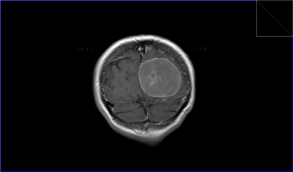
Meningiomas
EN

T2 AXIAL

FLAIR AXIAL

T1 CORONAL PRE CONTRAST

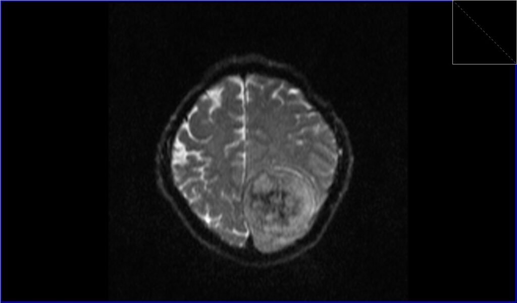
AXI DWI B0

AXI DWI B 1000

AXI DWI ADC

T1 CORONAL POST CONTRAST

T1 AXIAL POST CONTRAST


VN
Meningiomas are tumors that originate from the meninges, which are the layers of tissue covering the brain and spinal cord. These tumors are typically slow-growing and are usually benign (non-cancerous) in nature. However, some meningiomas can exhibit more aggressive behavior and rare cases can be malignant (cancerous).
T2 AXIAL
FLAIR AXIAL
T1 CORONAL PRE CONTRAST
AXI DWI B0
AXI DWI B 1000
AXI DWI ADC
T1 CORONAL POST CONTRAST
T1 AXIAL POST CONTRAST